Bone Cross Section Diagram : : Diagram with articular cartilage, marrow, spongy bone, medullary cavity, endosteum, diaphysis, and periosteum.. The cross section of this object is a triangle. Bone cross section for radius digital science on behance. Dinosaurs skeletons bones in soil layers vector. From wikimedia commons, the free media repository. A cross section of a human long bone.
Vector illustration scheme of bone cross section. Bone is found in the shafts of long bone and consists of various cylindrical units named as haversian system 47. They build the entire picture, improve your understanding, consolidate the information and facilitate recall. Cross section through middle metacarpal bones of vector. Vector illustration scheme of bone cross section.
Femur Cross-Section - Anatomy Pictures and Information from www.innerbody.com Two prominent grooves or sulci run along its length. Explaned distal and proximal epiphysis. Vector illustration scheme of bone cross section. For example, to read this diagram literally, since the cartilage can be seen inside the cutaway section of bone, it. The cross section of this object is a triangle. Histology sauropod vertebra picture of the week these pictures of this page are about:long bone cross section. As shown in figure 2. In a cross section of a bone we can see two types of bone tissue:
Spongy bone and compact bone.
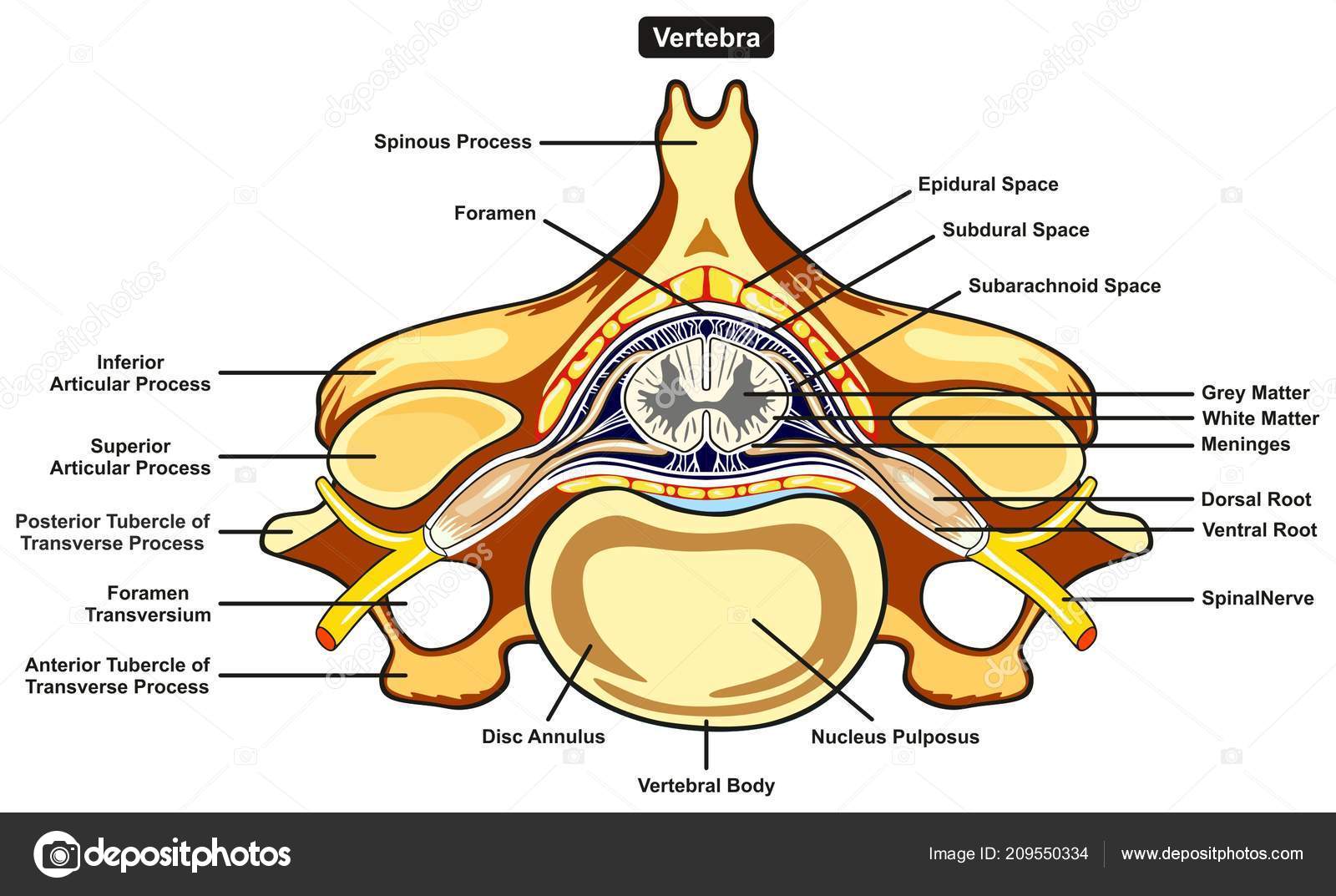
Spongy bone and compact bone. Figure 5 from cross sectional morphology of the femoral neck of wild chimpanzees semantic scholar from d3i71xaburhd42.cloudfront.net. Vector illustration scheme of bone cross section. Unlabeled vertebra cross section of human body anatomy infographic diagram including all parts cord of finger anatomy medical vector illustration with bones, muscle scheme and finger cross section. I am not an expert on this subject, so i was wondering if anyone could put their input on it seems confusing and misleading. Haz tu selección entre imágenes premium sobre bone cross section de la más alta calidad. The centroidal locations of common cross sections are well documented, so it is typically not necessary to calculate the location with the equations above. Bone cross section for radius digital science on behance. Cross section of bone diagram. Bone cross section diagram ipad folio cases. Vector illustration scheme of bone cross section. Cord spinal cross section spine cervical diagram education science anatomical anatomy atlas back body bone care column disc disease foramen fracture grey health healthcare healthy human illustration infographic injury matter medical nerve nervous pain part physiology poster process skeletal skeleton. Bone is found in the shafts of long bone and consists of various cylindrical units named as haversian system 47.
Diagram of a cross section of the coiled cochlea. Dinosaurs skeletons bones in soil layers vector. Bone cross section diagram card | zazzle. This is a short tutorial using blender 2.8 that shows how to create a bone cross section and using images to create the textures. Knee joint cross for basic medical education also vector.
Spinal Cord Cross Section Diagram Spinal Cord Cross ... from i.pinimg.com Explaned distal and proximal epiphysis. Spongy bone and compact bone. Bone cross section for radius digital science on behance. As shown in figure 2. Bone cross section diagram ipad folio cases. Diagram with articular cartilage, marrow, spongy bone, medullary cavity, endosteum, diaphysis, and periosteum. Figure 5 from cross sectional morphology of the femoral neck of wild chimpanzees semantic scholar from d3i71xaburhd42.cloudfront.net. Hope you enjoy and please.
There are trabeculae in spongy bone which gives its sponge like appearance.
Bone is found in the shafts of long bone and consists of various cylindrical units named as haversian system 47. Diagram with articular cartilage, marrow, spongy bone, medullary cavity, endosteum, diaphysis, and periosteum. Knee joint cross for basic medical education also vector. Cross section of the human retina. 12 photos of the cross section of human bone diagram. As with other tools applied to petroleum development. Cross section of bone diagram. 512 x 512 jpeg 27kb. From wikimedia commons, the free media repository. Diagram with articular cartilage, marrow, spongy bone, medullary cavity, endosteum, diaphysis, and periosteum. A cross section is the shape we get when cutting straight through an object. This is a short tutorial using blender 2.8 that shows how to create a bone cross section and using images to create the textures. It is like a view into the inside of something made by cutting through it.
Histology sauropod vertebra picture of the week these pictures of this page are about:long bone cross section. Jump to navigation jump to search. As with other tools applied to petroleum development. This is why anatomical position for the hand in medical diagrams is with the thumb pointing out instead of the more natural pointing in, so the radius and ulna are parallel instead of crossing over each other. Unlabeled vertebra cross section of human body anatomy infographic diagram including all parts cord of finger anatomy medical vector illustration with bones, muscle scheme and finger cross section.
Human body labeled | Labeled Vertebra Cross Section Human ... from st4.depositphotos.com Compact bone areas with numerous interconnecting cavities corresponding to. Knee joint cross for basic medical education also vector. Bone cross section for radius digital science on behance. Dinosaurs skeletons bones in soil layers vector. Haz tu selección entre imágenes premium sobre bone cross section de la más alta calidad. Bone on hand and foot diagram quiz. Spongy bone and compact bone. Diagram with articular cartilage, marrow, spongy bone, medullary cavity, endosteum, diaphysis, and periosteum.
Spinal cord spinal column anatomy information myvmc.
Unlabeled vertebra cross section of human body anatomy infographic diagram including all parts cord of finger anatomy medical vector illustration with bones, muscle scheme and finger cross section bone cross section. Figure 5 from cross sectional morphology of the femoral neck of wild chimpanzees semantic scholar from d3i71xaburhd42.cloudfront.net.


0 Komentar